In addition to their well-known role in regulating inflammatory process in the skin, sensory neurons are able to accelerate cell migration and have to be activated to induce a proper re-innervation of the scar.
Many factors ranging from the most benign to the most serious are the cause of formation of sores on the skin (injury, burn, surgical operation, pathology). These wounds are a possible source of entry of pathogens. Poor healing can lead to an alteration in the physiology of the skin.
It is therefore important to improve the healing process to restore the barrier function of the skin as quickly as possible, but also its other functionalities and sensoriality.
The healing process includes three major stages: the inflammatory phase, the tissue repair/proliferative phase itself and the maturation/remodeling phase. During the inflammatory phase, immune system cells will be recruited to the wound in order to clean it and keep it healthy. During the repair/proliferative phase a new connective tissue matrix is formed and keratinocytes migrate and proliferate in the wound to reform the epidermis and fill the wound. During the modeling phase cells and extra cellular matrice undergo a maturation process with the reorganization of tissues to form a fully healed tissue.
Sensory neurons being able to promote anti- and/or pro-inflammatory cytokines release by skin cells, targeting them with active ingredients is of interest for balancing the inflammatory process during wound healing. Furthermore, sensory neurons are able to accelerate cell migration and have to be activated to induce a proper re-innervation of the scar.
Neuron Experts has developed a model of co-culture of human sensory neurons and keratinocytes allowing to evaluate the ability of active ingredients to act on sensory neurons to modulate the wound healing process.
Cell System
- Humans iPS derived sensory neurons + skin cells (keratinocytes…)
Model of activation
- CGRP
- NGF
Endpoint Evaluation
- Percentage of cell migration in scar surface acquired at different time-points
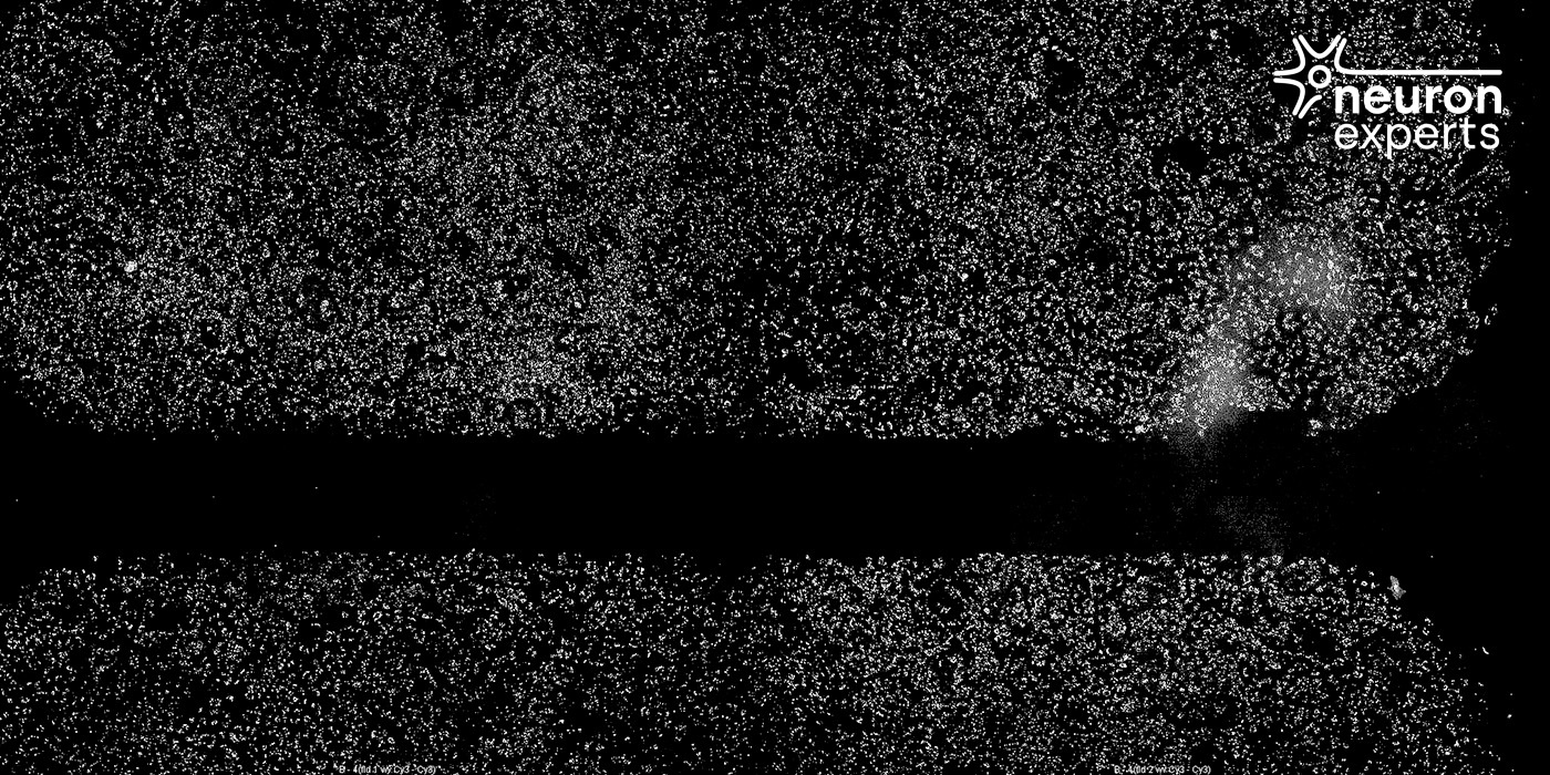
Human sensory neuron in coculture with keratinocytes in High-Throughput Wound Healing Assays (Ibidi plates), Cell viability probe (Cell Tracker TMCM-Dil Dye), T=0
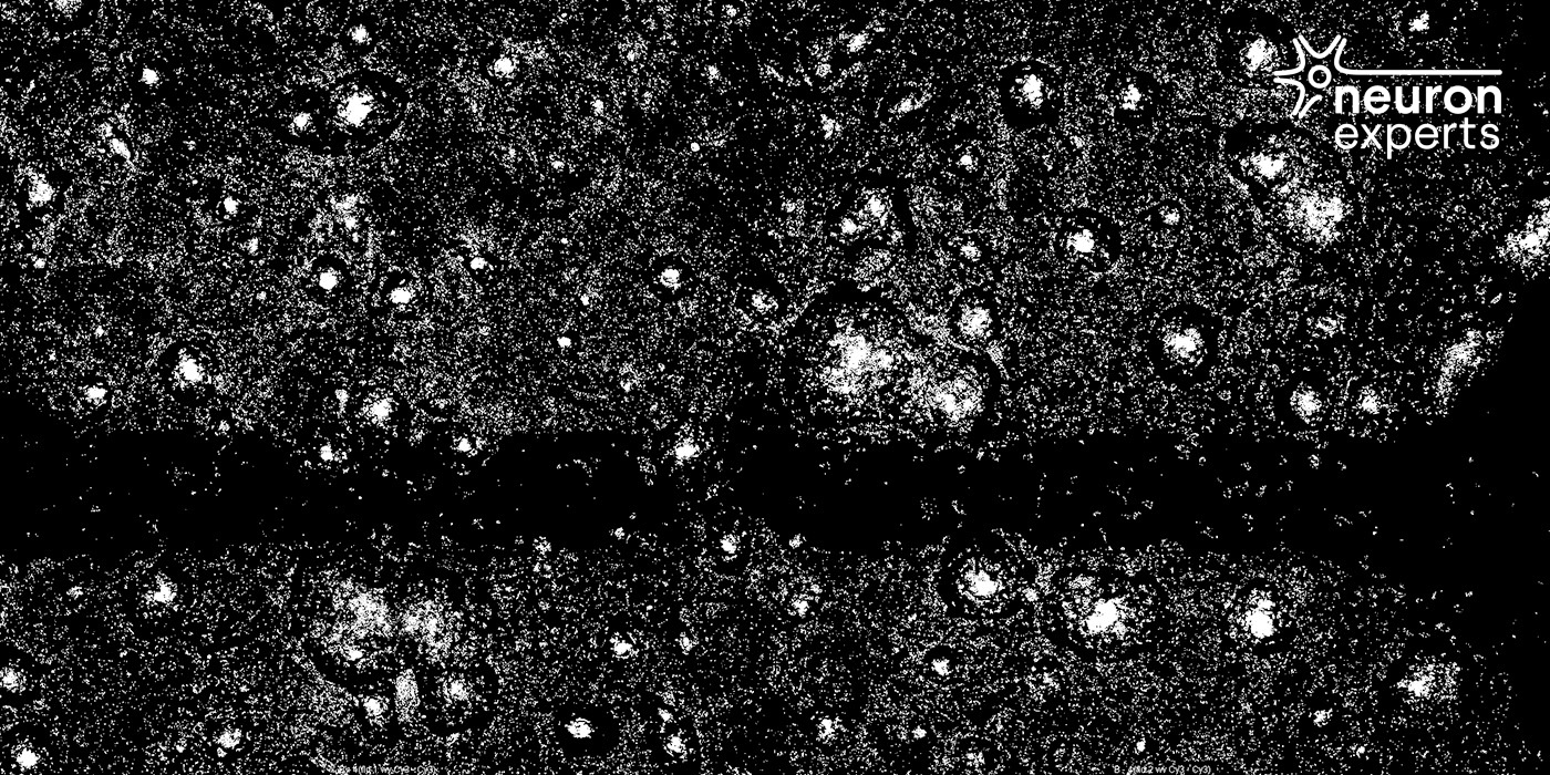
Human sensory neuron in coculture with keratinocytes in High-Throughput Wound Healing Assays (Ibidi plates), Cell viability probe (Cell Tracker TMCM-Dil Dye), T=30 hours
- Neurite detection in the scar surface
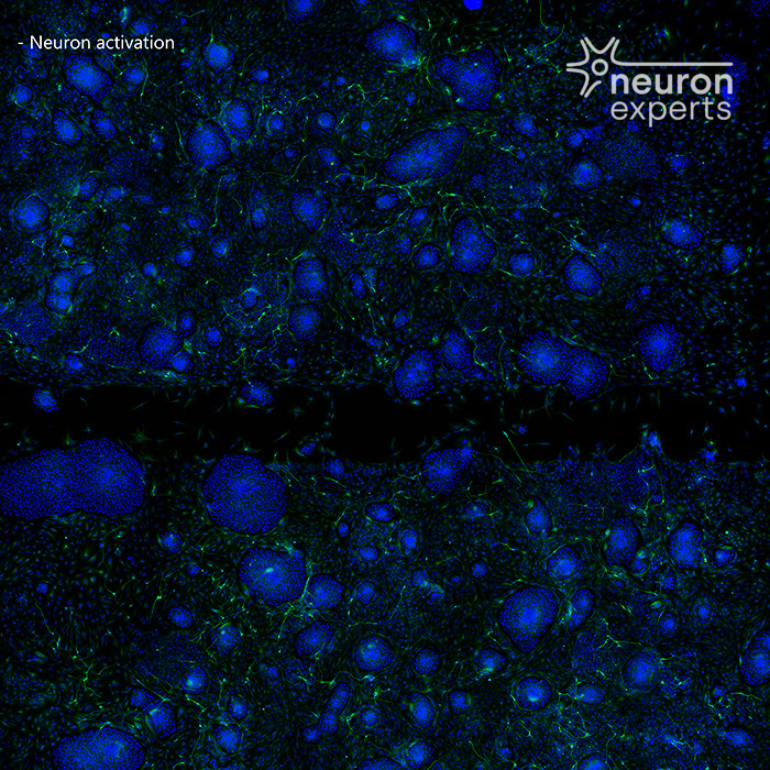
Human sensory neuron in coculture with keratinocytes in High-Throughput Wound Healing Assays (Ibidi plates), Green: β-tubulin; Blue: DAPI, T=0
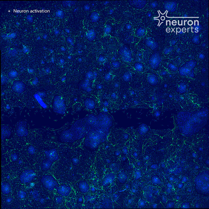
Human sensory neuron in coculture with keratinocytes in High-Throughput Wound Healing Assays (Ibidi plates), Green: β-tubulin; Blue: DAPI, T=30 hours