The pathophysiology of Alzheimer’s disease (AD) is characterized by chronic, progressive neurodegeneration. The precise aetiology of AD is still not fully clarified but is known to be complex and multifactorial. The neurodegeneration seen in AD involves a progressive accumulation of intracellular neurofibrillary tangles, extracellular parenchymal senile plaques, and cerebrovascular deposits; comprised of amyloid-β (Aβ) peptides.
Aβ peptide is a proteolytic product derived through sequential
proteolysis of amyloid precursor protein (APP), which occurs as a result of cleavage
by β-secretase and γ-secretase. Mutations in the cleavage of APP increase the
production of Aβ oligomers. The progressive accumulation of Aβ in the form of senile
plaques, which is one of the pathologic hallmarks of Alzheimer’s disease (AD), has
been recognized as one of the major causes of AD pathology by triggering
1-neurotoxicity, 2-oxidative damage, 3-inflammation.
Senile plaques are not only able to activate microglia but also to induce oxidative
stress by a release of H2O2 causing an alteration of axon membranes, an increase of
intracellular Ca2+ and Tau hyperphosphorylation that trigger neurofibrils formation,
decrease of microtubule attachment and axonal trafficking. The accumulation of Aβ in
synapses and synaptic mitochondria causes synaptic mitochondrial failure and
synaptic degeneration in Alzheimer’s disease (AD).
The most abundant Aβ peptide forms found in AD brain senile plaques are the 40 and
42 amino acid forms, the Aβ 1-42 having been ascribed as the main pathogenic form of
this peptide. Nevertheless, the number of senile plaques in a particular region of
the AD brain correlates poorly with the local extent of neuron death or synaptic
loss, or with cognitive impairment. Recent studies show a strong correlation between
the soluble Aβ oligomer (AβO) levels and the extent of synaptic loss and severity of
cognitive impairment.
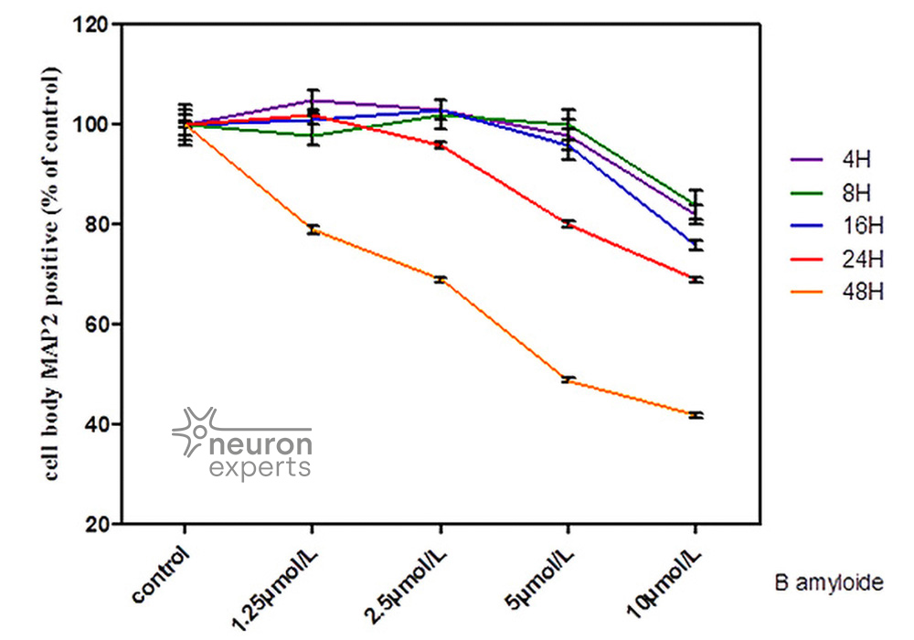
Neuron Experts developed an original method to generate with human β amyloid 1-42, a solution of AβO; forms of Aβ proved to be toxic.
Cell System
- Primary culture of cortical neurons
- Primary culture of hippocampal neurons
- Glial cells (astrocytes - microglial cells)
- Cortical or hippocampal neurons culture in Microfluidic Chamber
- Human IPS derivated cortical neurons
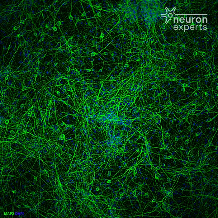
Primary culture of cortical neurons before 1-42 beta Amyloid Peptide oligomers injury
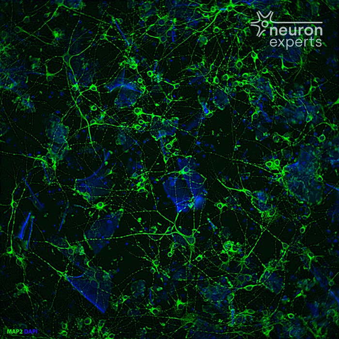
Primary culture of cortical neurons after (2) 1-42 beta Amyloid Peptide oligomers injury
Models of Intoxication
- Glutamate or NMDA injuries
- 1-42 beta Amyloid Peptide oligomers (A β O) injuries
- TNFα injuries
- Hypoxia
- Tau PFFs
Endpoint Evalution
- Neuronal death and apoptosis evaluation (Caspases 3 and 9)
- Loss of neurite (microtubule destabilisation)
- Hyperphosphorylation of Tau protein
- Synapses loss
- Oxidative stress
- Mitochondria injuries
- Autophagy evaluation (Lamp 2 - LC3b)
- Intracellular calcium quantification
- Release cytokines
- ....
Primary culture of cortical neurons after (2) 1-42 beta Amyloid Peptide oligomers injury